


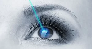
 Lasik – About the Procedure
Safest and best Lasik Eye Surgery & Treatment in Bangalore
LASIK(Laser Insitu Keratomilieusis)is a procedure where the need to use glasses or contact lenses is eliminated using an Excimer laser.
There are two steps in the LASIK procedure.First,the surgeon creates a micro-thin corneal flap ,which is lifted to expose the inner cornea for step two,tissue ablation by an excimer laser.The first step of making flap can be done either with ultrathin blade call Microkeratome(Blade LASIK) or with laser(Blade free LASIK).
The Femto second (FS) laser introduced the concept of blade-free LASIK,representing the first improvement to the procedure’s first step.Surgeons have found statistically and clinically significant differences in the vision patients achieve-better than 20/20 to 20/15 and even 20/12.5 -When the laser is used to make the corneal flap.The Femto second(FS) laser introduced the concept of blade-free LASIK,representing the first improvement to the procedure’s first step.Surgeons have found statistically and clinically significant…
Cataract surgery is the most frequently performed surgical procedure in the world. In conventional phacoemulsification cataract surgery, the surgeon uses hand held instruments to manually create a circular opening in the outer shell of the cataract. Next the surgeon uses ultrasound energy and instruments to break up and remove the cataract. Finally, the surgeon implants an artificial intraocular lens inside the remaining shell to replace the natural lens.
The aim of the surgery is to provide patients with the best possible post-operative vision. Cataract surgery outcomes are based on planning, managing and performing the surgery and the effective monitoring of the post-operative period.
At Prabha, you will benefit from the worlds’ best array of instruments and services backed with the experience of having performed over 125000 surgeries.
Our team of doctors is experienced operating on patients needing complex and combined cataract surgery with histories of Glaucoma (click here), Retinal problems (click here), Paediatric cataracts and in uveitic eyes (click here)
Surgical planning: At Prabha Eye Clinic benefit from the most accurate pre-surgical planning suite to ensure the best possible IOL and surgery method choice for your eye

Types of Contact Lenses
Many types of contact lenses are available. The usage of a particular type of contact lens is prescribed by the eye doctor. The following are the different types available:
Rigid or “hard” contacts were the first lenses; they were developed in the 1960’s. They are made of a type of plastic called PMMA (polymethyl methacrylate), which is very durable, but does not allow oxygen in the air to directly reach the cornea. When the eye blinks, the lens moves, which allows the oxygen dissolved in the tears to reach the cornea. Rigid lenses are the least comfortable type of contacts and are not really used anymore. However, some people still prefer them for their durability and lower cost.
These lenses are also known as “RGPs.” They are newer rigid or “hard” lenses made of plastics combined with other materials, such as silicone and fluoropolymers, which allow oxygen in the air to pass directly through the lens. For this reason, they are called “gas permeable”.
These lenses are made of plastic materials that incorporate water. The water makes them soft and flexible, as well as allowing oxygen to reach the cornea. Extended wear contact lenses: made of material designed to last 2-4 weeks. Most of the people prefer soft contact lenses due to the flexibility.
PMMA Lenses:
Gas-Permeable Lenses:
Soft Contact Lenses:
although generally more expensive, carry a lower infection risk.
correct moderate astigmatism. They are available in both rigid and soft materials.
Daily Disposable Lenses:
Toric Contact Lenses:
Contacts should be removed at bedtime due to risk of infection and risk of contact lens intolerance.
What is the Cost?
Usually the price of contact lens includes that of the lenses only. However it may include the cost of eye examination. In case a treatment not related to an eye condition is required, then the charges may be additional.
What Are the Risks?
PMMA lenses are more likely to scratch the cornea if the lens does not fit properly or if the lens is worn while sleeping. They are also more likely to slide off the cornea and become hidden under the lid. Rigid lenses traditionally had a reputation for “popping out” of the eye. New lens designs have minimized the chance of loosing a contact even during vigorous exercise. Rigid gas-permeable lenses and soft extended-wear contacts are the most likely to have protein build-up and cause lens-related allergies. Protein build-up results in discomfort, blurring and intolerance to the lenses. You will need special cleaning solutions to dissolve the protein.
Daily-wear lenses should never be worn as extended-wear lenses. Misuse can lead to temporary and even permanent damage to the cornea. People who wear any type of lens overnight have a greater chance of developing infections of the cornea. These infections are often due to poor cleaning and lens care. Improper over wearing of contact lenses can result in intolerance, leading to the inability to wear contact lenses.
Rigid gas-permeable or disposable lenses may be good choices for someone with allergies.
Who should not Wear Contact Lenses?
Contact lenses can be worn by people who require vision correction. But some people who have frequent eye infections, allergies, dry eye, and very dusty work environment cannot wear contact lenses due to the inability to handle and care for the lenses properly.
Are Contacts for You?
The requirement of contacts by people varies on:
Individual needs and expectations.
Patience and motivation during the initial adjustment period to contact lens wear.
Adhering to contact lens guidelines for wear, disinfecting and cleaning.
Diagnosis and treatment of conditions that may prevent contact lens wear.
How Do you Care for them?
Contact lenses must be properly cleaned and disinfected when you remove them to kill germs and prevent infections.
At the time you insert your contact lenses, you should thoroughly rinse the case with warm water and allow it to dry. All contact lens cases need frequent cleaning, including disposable lens cases.
Do not put your lens in your mouth and then in your eye.
Do not use homemade cleaning solutions, they have been linked to serious eye infections.
Do not attempt to sterilize disposable lenses – throw them away.
Do not mix different brands of solutions.
Any eye drops, even nonprescription ones, can interact with all types of contact lenses. Use the brand of solution prescribed by our doctor or check with the doctor before changing brands.
Wear your Lenses properly
Contact lenses must be properly cleaned and disinfected when you remove them to kill germs and prevent infections.
At the time you insert your contact lenses, you should thoroughly rinse the case with warm water and allow it to dry. All contact lens cases need frequent cleaning, including disposable lens cases.
Do not put your lens in your mouth and then in your eye.
Do not use homemade cleaning solutions, they have been linked to serious eye infections.
Do not attempt to sterilize disposable lenses – throw them away.
Do not mix different brands of solutions.
Any eye drops, even nonprescription ones, can interact with all types of contact lenses. Use the brand of solution prescribed by our doctor or check with the doctor before changing brands.
Call your Doctor when you Notice any of these Symptoms
Your eye is painful,
Your eye is red for more than two days,
You have discharge from your eye,
You have blurry vision,
Your eye feels scratchy.
ORTHO K LENS
Orthokeratology lenses are specially designed gas permeable [RGP] contact lenses used to alter the shape of the cornea in order to reduce or correct myopia [ short sight]. It can also be effective in low degrees of certain types of astigmatism. These lenses work best with a maximum of up to 1 diopter cylinder & -5 sphere. They are worn overnight and are removed on waking up in the morning. It takes about a month for visual correction to occur. Contact us for more details
ROSE K LENS
Rose K lenses are gas permeable lenses designed especially for keratoconus. These lenses are custom fit to each eye to correct myopia and astigmatism resulting from keratoconus.
SCLERAL LENS
Scleral contact lenses are large diameter gas permeable contact lenses for keratoconus and irregular corneas. They are also used in patients whose eyes are very sensitive to normal RGP contact lenses but require a more rigid lens for visual correction especially in higher degrees of astigmatism.
 With an increasing number of patients coming to us with complaints of dry eye and occupation related eye strain, we have now opened a dedicated clinic for Dry Eyes and Computer Vision Syndrome. This Clinic is open every day with prior appointment
Computer Use and Eyes
nstead of starring on the silver screen or playing outside, a great number of people spend the majority of their workday in front of a computer screen. By one estimate, nearly 90 percent of those people working at a video display terminal (VDT) experienced some form of vision problem as a result.
How do you Tell if Your Symptoms are Related to Your Computer Use?
VDT-related symptoms occur some time after you start work. As the workday progresses, your symptoms will become more acute. What are the symptoms that are related to computer use?
Difficulty focusing after working at a computer, with
blurry or double vision.
Eyestrain or eye fatigue
Headaches or backaches
Dry and/or irritated eyes
Neck stiffness or discomfort
After-images when looking away from the screen
Sensitivity to lighting
Muscle spasms
Are there Environmental Factors that could Affect your symptoms?
Bright lights in your peripheral field of vision could add to discomfort or reduced visual performance. Reflected light on your computer screen can decrease the contrast of screen characters and possibly force you to assume an awkward position to see around the glare.
The location of your screen could cause awkward positioning.
To determine the cause of your symptoms, you should visit your eye care professional. Before going to your appointment, however, take note of the environment in which your symptoms occur and at what times they are greatest. This will aid your doctor greatly in the diagnosis.
 We manage all types of glaucoma and provide comprehensive clinical examination including Gonioscopy, Tonometry and detailed Fundus evaluation. Investigative modalities that we have include Humphrey Visual Field Analyser, Optical Coherence Tomography, Pachymetry, Ultrasound Biomicroscopy and our newly acquired Anterion for anterior segment imaging.
Laser treatment modalities that we provide include, Nd YAG Laser for peripheral iridotomies, and capsulotomies. We also perform Trans-scleral and Endoscopic Cyclophotocoagulation, Cyclocryotherapy in advanced glaucoma. We offer a wide range of Surgical options from the most conventional to the latest, and include Trabeculectomy, Ahmed Glaucoma Implant and Minimally Invasive Glaucoma Surgeries such as IStent, Kahook Dual Blade(KDB) excisional goniotomy and Bent angle needle goniotomy(BANG). Specialised and complex cataract surgeries in eyes with previous glaucoma surgeries, Pseudoexfoliation, non-dilating pupils, traumatic eyes needing repair and special conditions like nanophthalmos, anterior segment dysgenesis are also done in addition to the standard combined cataract and Glaucoma surgical procedures.
We also extend our services to the pediatric population and offer clinical and surgical management of Glaucoma in newborn, infants and children. Goniotomy and Trabeculotomy are also performed. Continued patient education and availability of patient counselling services helps us in a providing a more holistic approach to patient-care
More Info on Glaucoma
About Glaucoma Surgery -will provide from new brochure text
General Information about Glaucomawill provide from new brochure text
Laser Treatment in Glaucoma –
Glaucoma laser surgery is usually done in a clinic and it is painless. During laser, a tiny but powerful beam of light is used to treat a part of the eye .

Lasik – About the Procedure
Safest and best Lasik Eye Surgery & Treatment in Bangalore
LASIK(Laser Insitu Keratomilieusis)is a procedure where the need to use glasses or contact lenses is eliminated using an Excimer laser.
There are two steps in the LASIK procedure.First,the surgeon creates a micro-thin corneal flap ,which is lifted to expose the inner cornea for step two,tissue ablation by an excimer laser.The first step of making flap can be done either with ultrathin blade call Microkeratome(Blade LASIK) or with laser(Blade free LASIK).
The Femto second (FS) laser introduced the concept of blade-free LASIK,representing the first improvement to the procedure’s first step.Surgeons have found statistically and clinically significant differences in the vision patients achieve-better than 20/20 to 20/15 and even 20/12.5 -When the laser is used to make the corneal flap.The Femto second(FS) laser introduced the concept of blade-free LASIK,representing the first improvement to the procedure’s first step.Surgeons have found statistically and clinically significant…
Types of Contact Lenses
Many types of contact lenses are available. The usage of a particular type of contact lens is prescribed by the eye doctor. The following are the different types available:
Rigid or “hard” contacts were the first lenses; they were developed in the 1960’s. They are made of a type of plastic called PMMA (polymethyl methacrylate), which is very durable, but does not allow oxygen in the air to directly reach the cornea. When the eye blinks, the lens moves, which allows the oxygen dissolved in the tears to reach the cornea. Rigid lenses are the least comfortable type of contacts and are not really used anymore. However, some people still prefer them for their durability and lower cost.
These lenses are also known as “RGPs.” They are newer rigid or “hard” lenses made of plastics combined with other materials, such as silicone and fluoropolymers, which allow oxygen in the air to pass directly through the lens. For this reason, they are called “gas permeable”.
These lenses are made of plastic materials that incorporate water. The water makes them soft and flexible, as well as allowing oxygen to reach the cornea. Extended wear contact lenses: made of material designed to last 2-4 weeks. Most of the people prefer soft contact lenses due to the flexibility.
PMMA Lenses:
Gas-Permeable Lenses:
Soft Contact Lenses:
although generally more expensive, carry a lower infection risk.
correct moderate astigmatism. They are available in both rigid and soft materials.
Daily Disposable Lenses:
Toric Contact Lenses:
Contacts should be removed at bedtime due to risk of infection and risk of contact lens intolerance.
What is the Cost?
Usually the price of contact lens includes that of the lenses only. However it may include the cost of eye examination. In case a treatment not related to an eye condition is required, then the charges may be additional.
What Are the Risks?
PMMA lenses are more likely to scratch the cornea if the lens does not fit properly or if the lens is worn while sleeping. They are also more likely to slide off the cornea and become hidden under the lid. Rigid lenses traditionally had a reputation for “popping out” of the eye. New lens designs have minimized the chance of loosing a contact even during vigorous exercise. Rigid gas-permeable lenses and soft extended-wear contacts are the most likely to have protein build-up and cause lens-related allergies. Protein build-up results in discomfort, blurring and intolerance to the lenses. You will need special cleaning solutions to dissolve the protein.
Daily-wear lenses should never be worn as extended-wear lenses. Misuse can lead to temporary and even permanent damage to the cornea. People who wear any type of lens overnight have a greater chance of developing infections of the cornea. These infections are often due to poor cleaning and lens care. Improper over wearing of contact lenses can result in intolerance, leading to the inability to wear contact lenses.
Rigid gas-permeable or disposable lenses may be good choices for someone with allergies.
Who should not Wear Contact Lenses?
Contact lenses can be worn by people who require vision correction. But some people who have frequent eye infections, allergies, dry eye, and very dusty work environment cannot wear contact lenses due to the inability to handle and care for the lenses properly.
Are Contacts for You?
The requirement of contacts by people varies on:
Individual needs and expectations.
Patience and motivation during the initial adjustment period to contact lens wear.
Adhering to contact lens guidelines for wear, disinfecting and cleaning.
Diagnosis and treatment of conditions that may prevent contact lens wear.
How Do you Care for them?
Contact lenses must be properly cleaned and disinfected when you remove them to kill germs and prevent infections.
At the time you insert your contact lenses, you should thoroughly rinse the case with warm water and allow it to dry. All contact lens cases need frequent cleaning, including disposable lens cases.
Do not put your lens in your mouth and then in your eye.
Do not use homemade cleaning solutions, they have been linked to serious eye infections.
Do not attempt to sterilize disposable lenses – throw them away.
Do not mix different brands of solutions.
Any eye drops, even nonprescription ones, can interact with all types of contact lenses. Use the brand of solution prescribed by our doctor or check with the doctor before changing brands.
Wear your Lenses properly
Contact lenses must be properly cleaned and disinfected when you remove them to kill germs and prevent infections.
At the time you insert your contact lenses, you should thoroughly rinse the case with warm water and allow it to dry. All contact lens cases need frequent cleaning, including disposable lens cases.
Do not put your lens in your mouth and then in your eye.
Do not use homemade cleaning solutions, they have been linked to serious eye infections.
Do not attempt to sterilize disposable lenses – throw them away.
Do not mix different brands of solutions.
Any eye drops, even nonprescription ones, can interact with all types of contact lenses. Use the brand of solution prescribed by our doctor or check with the doctor before changing brands.
Call your Doctor when you Notice any of these Symptoms
Your eye is painful,
Your eye is red for more than two days,
You have discharge from your eye,
You have blurry vision,
Your eye feels scratchy.
ORTHO K LENS
Orthokeratology lenses are specially designed gas permeable [RGP] contact lenses used to alter the shape of the cornea in order to reduce or correct myopia [ short sight]. It can also be effective in low degrees of certain types of astigmatism. These lenses work best with a maximum of up to 1 diopter cylinder & -5 sphere. They are worn overnight and are removed on waking up in the morning. It takes about a month for visual correction to occur. Contact us for more details
ROSE K LENS
Rose K lenses are gas permeable lenses designed especially for keratoconus. These lenses are custom fit to each eye to correct myopia and astigmatism resulting from keratoconus.
SCLERAL LENS
Scleral contact lenses are large diameter gas permeable contact lenses for keratoconus and irregular corneas. They are also used in patients whose eyes are very sensitive to normal RGP contact lenses but require a more rigid lens for visual correction especially in higher degrees of astigmatism.


Disorders Treated
Diabetic retinopathy
Diabetic retinopathy is a complication of diabetes and a leading cause of blindness. It occurs whendiabetes damages the tiny blood vessels inside the retina which comprises the light-sensitive tissueat the back of the eye. A healthy retina is necessary for good vision. If you have diabetic retinopathy,at first you may notice no changes to your vision. But over time, diabetic retinopathy can worsenand cause vision loss. Timely intervention in the form of laser , intraocular injections or apt surgical treatment will arrest the progression of the disease, along with adequate diabetic systemic control.
Age related macular degeneration
In macular degeneration, the light-sensing cells of the macula malfunction and may over time ceaseto work. Macular degeneration occurs most often in people over 60 years old, in which case it iscalled Age Related Macular Degeneration (ARMD). ARMD can be “dry” or “wet” depending on thedegree of damage. While dry ARMD can be observed – the wet type will need timely intraocular injections aimed at halting the disease progression.
Flashes and floaters
Light flashes are sometimes caused by mechanical stimulation of the retina. A variety of conditionscan cause it, including posterior vitreous separation and retinal tears. People who have a high minusrefractive error or in other words high myopia are most prone to developing retinal thinning holesand tears. A regular follow up with the retinal specialist will help in picking up the disease in its early stages.
Floaters are black coloured small objects that are seen in the field of view of the patient’s eyes.Sudden onset of these floaters can sometimes indicate retinal tear / bleeding into the cavity of the eye and has to be consulted with the retina specialist on an emergent basis.
Retinal detachment
Retinal detachments often develop in eyes with retinas weakened by a hole or tear. This allows fluidto seep underneath the retina , weakening the attachment so that the retina becomes detached likewallpaper peeling off a damp wall. When detached, the retina cannot form a clear picture and the
vision becomes blurred and dim. The retinal tissue starts to lose its functionality within 90 minutesof detachment. Hence these symptoms should be brought to the notice of the retina specialist atthe earliest so that a timely laser or surgical intervention can restore the vision to the best possible extent.
Retinal arterial blocks
A retinal artery occlusion occurs when the central retinal artery or one of its branches gets blocked.This blockage is typically caused by a tiny embolus (blood clot) in the bloodstream. The occlusiondecreases the oxygen supply to the area of the retina nourished by the affected artery, causingpermanent vision loss. Patients usually have a sudden loss of vision – typically when they wake upfrom sleep. Retinal artery obstruction is comparable to a stroke in the eye. The damage can be quitesevere, depending on the extent to which the blood flow has been disrupted.People withco-morbidities like diabetes, hypertension , underlying blood disorders are all prone to developing this condition.
Retinal venous blocks
Central and branch retinal venous occlusions occur when there is a congestion to blood flow and anincrease in backpressure on the central retinal vein. It causes a variable degree of visual loss andcan be easily diagnosed by a retinal examination. It is commonly seen in hypertension and diabetes.Sometimes it can be seen in people with blood clotting disorders. An early OCT scan of the retinacan help the retina specialist pick up a macular edema due to the occlusion and can be treated with intraocular injections aimed to salvage the vision to the best possible extent.
The eye is an important sense organ. It is made up of 3 coats. The outer coat is the cornea and sclera. The middle coat is the uveal tract and the inner coat is the retina. The uveal tract consists of 3 parts. The iris is the visible part of the uvea and gives color to the eye (blue, black, brown). The ciliary body is the middle layer of the uvea which manufactures the fluid inside the eye. The choroid is the posterior layer of the uvea which is rich in blood vessels.
What is Uveitis?
Inflammation or swelling in any of the part of the uveal tract is called uveitis. Depending on the location of inflammation, it is called anterior uveitis (iris and ciliary body is involved), intermediate uveitis (ciliary body and the vitreous is involved), posterior uveitis (choroids and retina involved), panuveitisd (all layers of the uveal tract are involved).
What Causes Uveitis?
Causes of Uveitis are many.
It may result from an infection (bacteria like tuberculosis, viruses like herpes, parasites like toxoplasmosis)
It may also be related to an autoimmune disease (with or without involvement of other parts of the body). This essentially is when our immune system recognizes a part of our own body as foreign and elicits a reaction in the form of uveitis. The uveitis may be the presenting manifestation of an underlying systemic disease.
It can occur as a result of injury to the eye.
It can occur due to causes within the eye like cataract and retinal detachment
Sometimes the cause is not known at the first instance. As we follow-up the patient over a period of time, we may be able to pick up new clues or signs suggestive of a systemic disease. In such cases, they are labeled as idiopathic (cause not known) at the initial visit.
What are the Symptoms of Uveitis?
Depending on the structure involved, the symptoms can vary from
Pain
Redness
Inability to see light (photophobia)
Black spots moving in front of the eye
Blurring or reduction in vision
How is an Ophthalmic Evaluation Done in a Patient with Uveitis?
An initial examination of the eye will be done. This includes the recording of the visual acuity by reading a chart (to check if the vision has decreased), slit lamp examination; (in which a narrow beam of light is shone into the eye so that a magnifying lens can closely examine the highlighted portion of the eye). Intraocular pressure recording (a painless test done by a tonometer); fundus examination (in which the pupil is widened so that the ophthalmologist can look into the eye and see structures in the back of the eye).
Because uveitis can be caused by so many different things (infections/autoimmune diseases) diagnosis may require detailed laboratory investigations and a referral to a uveitis specialist.
What Does a Uveitis Specialist Do?
A uveitis specialist is an ophthalmologist with a special, specific interest in uveitis and who has become proficient in the diagnosis and management of patients with uveitis. He or she has received adequate training in the field of uveitis.
A detailed medical history is elicited. As uveitis can occur due to infection or an underlying autoimmune disease, it is necessary to discover and treat these conditions as well. The results obtained in the routine laboratory tests and or imaging will be analyzed by the uveitis specialist. Many times a repeat in serologic testing may be needed. Specialized imaging of the eye and its structures may be required. Specialized blood tests related to the immune system may also be ordered to obtain a diagnosis. Depending on the type of uveitis, a sample of the fluid from the eye may also be sent to obtain a diagnosis by molecular diagnostic techniques. As uveitis may be related to a systemic disease, a cross consultation with a rheumatologist, a pulmonologist or a physician may also be done.
Why is Early Diagnosis of Uveitis Important?
Uveitis is the 3rd leading cause of blindness. Inflammation of the uvea can affect the cornea, retina, sclera and other vital parts of the body. Inflammation inside the eye is a medical emergency because, untreated, it will lead to vision loss. Damage to the eye is preventable if detected early and treated adequately.
How is Uveitis Treated?
The treatment of uveitis aims to achieve the following:
Relief of pain and discomfort (when present)
To prevent sight loss due to the disease or its complications
To treat the cause of the disease where possible.
Medical treatment of uveitis must be aggressive to prevent glaucoma, to prevent scarring of the structures inside the eye and to prevent possible blindness.
Different medications are used to control the original cause of uveitis, if known and to minimize the inflammation itself. Eye drops especially steroids (to reduce inflammation and pain) and pupil dilators (to widen the pupil and relax the muscles within the eye) are the main medications used to treat uveitis. For deeper inflammations, oral medicines eye injections or intravenous injections of steroids may be necessary. More severe cases of uveitis and those intolerant to steroids may require treatment with immunosuppressive agents. Complications such as glaucoma, cataract or new blood vessel formation (neovascularisation) also may need treatment in the course of the disease. If complications are advanced, conventional surgery or laser surgery may be necessary.
What is the Expected Duration of Treatment?
The duration of treatment varies from person to person and also depends on the type and cause of uveitis. Simple forms of uveitis, for example, may respond to treatment within days and may not recur. Chronic (long term, recurring) forms of uveitis that threaten vision can be very difficult to cure and require persistence on the part of the treating physician(s) and patient. The length of time required to get the disease into a durable remission on steroids or immunosuppressants is difficult to quantify and is very individualistic but the minimum of 2 years is a reasonable estimate. With appropriate, targeted treatment, most people with uveitis will become well controlled and progress to emission. Once in remission from uveitis, you should expect to have regular follow-up visits to our doctor to make sure that the disease remains in remission.
You have to Help to Save Your Sight.
Find support and encouragement from your family, friends and others. Sometimes it helps to talk to people who have experienced the same thing. It can help you to discuss side effects, share ways to remember your medicines and celebrate getting your uveitis under control. Unfortunately, there are a few people who will lose vision despite commitment to working with their eye doctor and following their treatment plan.
The future holds great promise for uveitis. New medicines and treatments continue to be developed. In the meantime, take heart in knowing that we are doing everything possible to treat your condition. The doctor/patient team approach, support from others and promising scientific discoveries will help you look forward towards a bright future.
 Neuro Ophthalmologists at Prabha Eye Clinic treat patients who have reduced vision, blind spot owing to disorders of the optic nerve and its connections to the brain leading to subnormal vision or field loss. Many conditions which can be life threatening can be diagnosed by eye examination by neuro ophthalmologist.
They also evaluate and treat any condition in which there may be a brain abnormality causing a disturbance of vision, misalignment of the eyes, or abnormal eye movements.
Our specialists also provide evaluation of focal dystonia, blepharospasm and hemi facial spasm, as well as botulinum toxin treatments for these conditions.
Optic Neuritis
Optic neuritis is an inflammation of the optic nerve. The optic nerve allows you to see by carrying images from your retina to your brain. The optic nerve resembles that of electrical wires or nerve fibers. Each wire carries a part of the visual information to the brain. If some or all of the nerve fibers become inflamed and do not function properly, vision becomes blurred. With optic neuritis, the optic nerve becomes swollen and the nerve fibers do not work properly. Vision can range from near normal to very poor depending on the number of inflamed nerve fibers.
Symptoms
Blurred vision in one or both eyes (especially after exercising or taking a hot bath).
Dim vision (as if the lights were turned down).
Abnormal color vision (dull and faded colors).
Pain behind the eye, particularly when moving the eyes.
The symptoms described above may not necessarily mean that you have optic neuritis. However, if you experience one or more of these symptoms, contact your eye doctor for a complete exam.
Treatment
Optic neuritis usually occurs suddenly. If you experience any of the symptoms listed above, call your ophthalmologist. By looking in the back of your eye with an instrument called the ophthalmoscope, ophthalmologist can see any optic nerve swelling. Optic neuritis may be confused with other causes of poor vision. Other tests such as color vision, side vision, and the reaction of the pupil to light may be performed. Ultrasound or magnetic scanning or visual brainwave recordings may be needed.
Optic Atrophy
Optic Atrophy means the loss of some or most of the nerve fibers in the optic nerve. (Resulting in severe visual loss). Many diseases and disorders can lead to optic atrophy or damage to one or both optic nerves. Optic atrophy can occur in people where the optic nerve or nerves did not develop properly. It may also result from inflammation of the optic nerve or from glaucoma when the pressure inside the eye remains too high. In unusual cases, poisons, vitamin deficiencies, or tumors may be responsible. Most commonly, optic atrophy simply occurs without a known or proven cause.
Symptoms
Blurred vision.
Abnormal side vision.
Abnormal color vision.
Poor constriction of the pupil in light
Decreased brightness in one eye relative to the other.
The symptoms described above may not necessarily mean that you have optic neuritis / optic neuropathy. However, if you experience one or more of these symptoms, contact your eye doctor for a complete exam.
Treatment
The optic nerve enters the back of the eye where it appears as a small disc, which your ophthalmologist can examine by looking through the pupil of your eye with a special instrument called an ophthalmoscope. If optic atrophy is present, this small disc will appear pale or white, indicating loss of nerve fibers. If you show some of the symptoms listed above, your ophthalmologist may decide to perform additional tests. Unfortunately, there is no effective treatment for optic atrophy. Once the nerve fibers in the optic nerve are lost they never heal or grow back. Therefore, the best defence is an early diagnosis because if the cause can be found and corrected, further damage can be prevented.
Adult Strabismus
Strabismus is a defect in the eyes wherein the eyes point in two different directions. The exact cause is not yet understood. Our strabismus patients receive comprehensive diagnosis and management of their ocular misalignment and diplopia. The services of certified orthoptists are important components of the program, offering special eye measurement tests as well as treatment with glasses, prisms, Bangerter Foils, and/or muscle exercises as required.
The treatment for Strabismus varies accordingly to the type of squint.
Strabismus can be either due to any craniomotor nerve palsies, any mechanical causes like fracture or bones of orbit, thyroid eye disease, neuro-muscular problems like myasthenia or due sensory strabismus due to poor vision.
The different types of treatment are:
Spectacles which is used to correct sight problems.
Occlusion – patching the good eye to encourage the weaker eye to be used.
Surgery is done in cases of congenital squints with other forms of treatment. During surgery a small incision is made in the tissue covering the eye to reach the eye muscles the eye muscles are removed from the wall of the eye and repositioned during the surgery, depending on which direction the eye is turning. Sometimes, it may be necessary to perform surgery on one or both eyes. Surgery in children in performed under general anesthesia. Migraine and other types of headache: Our neuro-ophthalmologists are trained in diagnosing and treating various types of headaches in children and in adults. A detailed history is taken and appropriate investigations are advised to know the root cause for headache and is treated in association with neurologists.
 Comprehensive fully automated clinical lab with quality bench marks certified by CMC Vellore’s External Quality System. The lab offers bio chemistry, immunochemistry, serology and ocular histo pathology services.
Comprehensive diagnostics services offered in association with Indias best lab.
 Comprehensive treatment and corrective surgical procedures are offered for
Droopy & tired eyes
Eyelid bags ( Blepharoplasty )
Wrinkles & deep expression lines (Botox treatment )
Dark circles
Aging eye and facial skin changes
Fat deposit around the eyes (Xanthelesma )
Treatment options available include
Ptosis surgery
Brow lift
Blepharoplasty (upper & lower)
Eyelid & facial skin tightening
Chemical peels, Medicated cosmeceuticals for circles & facial pigmentation
Disorders Treated
Diabetic retinopathy
Diabetic retinopathy is a complication of diabetes and a leading cause of blindness. It occurs whendiabetes damages the tiny blood vessels inside the retina which comprises the light-sensitive tissueat the back of the eye. A healthy retina is necessary for good vision. If you have diabetic retinopathy,at first you may notice no changes to your vision. But over time, diabetic retinopathy can worsenand cause vision loss. Timely intervention in the form of laser , intraocular injections or apt surgical treatment will arrest the progression of the disease, along with adequate diabetic systemic control.
Age related macular degeneration
In macular degeneration, the light-sensing cells of the macula malfunction and may over time ceaseto work. Macular degeneration occurs most often in people over 60 years old, in which case it iscalled Age Related Macular Degeneration (ARMD). ARMD can be “dry” or “wet” depending on thedegree of damage. While dry ARMD can be observed – the wet type will need timely intraocular injections aimed at halting the disease progression.
Flashes and floaters
Light flashes are sometimes caused by mechanical stimulation of the retina. A variety of conditionscan cause it, including posterior vitreous separation and retinal tears. People who have a high minusrefractive error or in other words high myopia are most prone to developing retinal thinning holesand tears. A regular follow up with the retinal specialist will help in picking up the disease in its early stages.
Floaters are black coloured small objects that are seen in the field of view of the patient’s eyes.Sudden onset of these floaters can sometimes indicate retinal tear / bleeding into the cavity of the eye and has to be consulted with the retina specialist on an emergent basis.
Retinal detachment
Retinal detachments often develop in eyes with retinas weakened by a hole or tear. This allows fluidto seep underneath the retina , weakening the attachment so that the retina becomes detached likewallpaper peeling off a damp wall. When detached, the retina cannot form a clear picture and the
vision becomes blurred and dim. The retinal tissue starts to lose its functionality within 90 minutesof detachment. Hence these symptoms should be brought to the notice of the retina specialist atthe earliest so that a timely laser or surgical intervention can restore the vision to the best possible extent.
Retinal arterial blocks
A retinal artery occlusion occurs when the central retinal artery or one of its branches gets blocked.This blockage is typically caused by a tiny embolus (blood clot) in the bloodstream. The occlusiondecreases the oxygen supply to the area of the retina nourished by the affected artery, causingpermanent vision loss. Patients usually have a sudden loss of vision – typically when they wake upfrom sleep. Retinal artery obstruction is comparable to a stroke in the eye. The damage can be quitesevere, depending on the extent to which the blood flow has been disrupted.People withco-morbidities like diabetes, hypertension , underlying blood disorders are all prone to developing this condition.
Retinal venous blocks
Central and branch retinal venous occlusions occur when there is a congestion to blood flow and anincrease in backpressure on the central retinal vein. It causes a variable degree of visual loss andcan be easily diagnosed by a retinal examination. It is commonly seen in hypertension and diabetes.Sometimes it can be seen in people with blood clotting disorders. An early OCT scan of the retinacan help the retina specialist pick up a macular edema due to the occlusion and can be treated with intraocular injections aimed to salvage the vision to the best possible extent.
The eye is an important sense organ. It is made up of 3 coats. The outer coat is the cornea and sclera. The middle coat is the uveal tract and the inner coat is the retina. The uveal tract consists of 3 parts. The iris is the visible part of the uvea and gives color to the eye (blue, black, brown). The ciliary body is the middle layer of the uvea which manufactures the fluid inside the eye. The choroid is the posterior layer of the uvea which is rich in blood vessels.
What is Uveitis?
Inflammation or swelling in any of the part of the uveal tract is called uveitis. Depending on the location of inflammation, it is called anterior uveitis (iris and ciliary body is involved), intermediate uveitis (ciliary body and the vitreous is involved), posterior uveitis (choroids and retina involved), panuveitisd (all layers of the uveal tract are involved).
What Causes Uveitis?
Causes of Uveitis are many.
It may result from an infection (bacteria like tuberculosis, viruses like herpes, parasites like toxoplasmosis)
It may also be related to an autoimmune disease (with or without involvement of other parts of the body). This essentially is when our immune system recognizes a part of our own body as foreign and elicits a reaction in the form of uveitis. The uveitis may be the presenting manifestation of an underlying systemic disease.
It can occur as a result of injury to the eye.
It can occur due to causes within the eye like cataract and retinal detachment
Sometimes the cause is not known at the first instance. As we follow-up the patient over a period of time, we may be able to pick up new clues or signs suggestive of a systemic disease. In such cases, they are labeled as idiopathic (cause not known) at the initial visit.
What are the Symptoms of Uveitis?
Depending on the structure involved, the symptoms can vary from
Pain
Redness
Inability to see light (photophobia)
Black spots moving in front of the eye
Blurring or reduction in vision
How is an Ophthalmic Evaluation Done in a Patient with Uveitis?
An initial examination of the eye will be done. This includes the recording of the visual acuity by reading a chart (to check if the vision has decreased), slit lamp examination; (in which a narrow beam of light is shone into the eye so that a magnifying lens can closely examine the highlighted portion of the eye). Intraocular pressure recording (a painless test done by a tonometer); fundus examination (in which the pupil is widened so that the ophthalmologist can look into the eye and see structures in the back of the eye).
Because uveitis can be caused by so many different things (infections/autoimmune diseases) diagnosis may require detailed laboratory investigations and a referral to a uveitis specialist.
What Does a Uveitis Specialist Do?
A uveitis specialist is an ophthalmologist with a special, specific interest in uveitis and who has become proficient in the diagnosis and management of patients with uveitis. He or she has received adequate training in the field of uveitis.
A detailed medical history is elicited. As uveitis can occur due to infection or an underlying autoimmune disease, it is necessary to discover and treat these conditions as well. The results obtained in the routine laboratory tests and or imaging will be analyzed by the uveitis specialist. Many times a repeat in serologic testing may be needed. Specialized imaging of the eye and its structures may be required. Specialized blood tests related to the immune system may also be ordered to obtain a diagnosis. Depending on the type of uveitis, a sample of the fluid from the eye may also be sent to obtain a diagnosis by molecular diagnostic techniques. As uveitis may be related to a systemic disease, a cross consultation with a rheumatologist, a pulmonologist or a physician may also be done.
Why is Early Diagnosis of Uveitis Important?
Uveitis is the 3rd leading cause of blindness. Inflammation of the uvea can affect the cornea, retina, sclera and other vital parts of the body. Inflammation inside the eye is a medical emergency because, untreated, it will lead to vision loss. Damage to the eye is preventable if detected early and treated adequately.
How is Uveitis Treated?
The treatment of uveitis aims to achieve the following:
Relief of pain and discomfort (when present)
To prevent sight loss due to the disease or its complications
To treat the cause of the disease where possible.
Medical treatment of uveitis must be aggressive to prevent glaucoma, to prevent scarring of the structures inside the eye and to prevent possible blindness.
Different medications are used to control the original cause of uveitis, if known and to minimize the inflammation itself. Eye drops especially steroids (to reduce inflammation and pain) and pupil dilators (to widen the pupil and relax the muscles within the eye) are the main medications used to treat uveitis. For deeper inflammations, oral medicines eye injections or intravenous injections of steroids may be necessary. More severe cases of uveitis and those intolerant to steroids may require treatment with immunosuppressive agents. Complications such as glaucoma, cataract or new blood vessel formation (neovascularisation) also may need treatment in the course of the disease. If complications are advanced, conventional surgery or laser surgery may be necessary.
What is the Expected Duration of Treatment?
The duration of treatment varies from person to person and also depends on the type and cause of uveitis. Simple forms of uveitis, for example, may respond to treatment within days and may not recur. Chronic (long term, recurring) forms of uveitis that threaten vision can be very difficult to cure and require persistence on the part of the treating physician(s) and patient. The length of time required to get the disease into a durable remission on steroids or immunosuppressants is difficult to quantify and is very individualistic but the minimum of 2 years is a reasonable estimate. With appropriate, targeted treatment, most people with uveitis will become well controlled and progress to emission. Once in remission from uveitis, you should expect to have regular follow-up visits to our doctor to make sure that the disease remains in remission.
You have to Help to Save Your Sight.
Find support and encouragement from your family, friends and others. Sometimes it helps to talk to people who have experienced the same thing. It can help you to discuss side effects, share ways to remember your medicines and celebrate getting your uveitis under control. Unfortunately, there are a few people who will lose vision despite commitment to working with their eye doctor and following their treatment plan.
The future holds great promise for uveitis. New medicines and treatments continue to be developed. In the meantime, take heart in knowing that we are doing everything possible to treat your condition. The doctor/patient team approach, support from others and promising scientific discoveries will help you look forward towards a bright future.
Neuro Ophthalmologists at Prabha Eye Clinic treat patients who have reduced vision, blind spot owing to disorders of the optic nerve and its connections to the brain leading to subnormal vision or field loss. Many conditions which can be life threatening can be diagnosed by eye examination by neuro ophthalmologist.
They also evaluate and treat any condition in which there may be a brain abnormality causing a disturbance of vision, misalignment of the eyes, or abnormal eye movements.
Our specialists also provide evaluation of focal dystonia, blepharospasm and hemi facial spasm, as well as botulinum toxin treatments for these conditions.
Optic Neuritis
Optic neuritis is an inflammation of the optic nerve. The optic nerve allows you to see by carrying images from your retina to your brain. The optic nerve resembles that of electrical wires or nerve fibers. Each wire carries a part of the visual information to the brain. If some or all of the nerve fibers become inflamed and do not function properly, vision becomes blurred. With optic neuritis, the optic nerve becomes swollen and the nerve fibers do not work properly. Vision can range from near normal to very poor depending on the number of inflamed nerve fibers.
Symptoms
Blurred vision in one or both eyes (especially after exercising or taking a hot bath).
Dim vision (as if the lights were turned down).
Abnormal color vision (dull and faded colors).
Pain behind the eye, particularly when moving the eyes.
The symptoms described above may not necessarily mean that you have optic neuritis. However, if you experience one or more of these symptoms, contact your eye doctor for a complete exam.
Treatment
Optic neuritis usually occurs suddenly. If you experience any of the symptoms listed above, call your ophthalmologist. By looking in the back of your eye with an instrument called the ophthalmoscope, ophthalmologist can see any optic nerve swelling. Optic neuritis may be confused with other causes of poor vision. Other tests such as color vision, side vision, and the reaction of the pupil to light may be performed. Ultrasound or magnetic scanning or visual brainwave recordings may be needed.
Optic Atrophy
Optic Atrophy means the loss of some or most of the nerve fibers in the optic nerve. (Resulting in severe visual loss). Many diseases and disorders can lead to optic atrophy or damage to one or both optic nerves. Optic atrophy can occur in people where the optic nerve or nerves did not develop properly. It may also result from inflammation of the optic nerve or from glaucoma when the pressure inside the eye remains too high. In unusual cases, poisons, vitamin deficiencies, or tumors may be responsible. Most commonly, optic atrophy simply occurs without a known or proven cause.
Symptoms
Blurred vision.
Abnormal side vision.
Abnormal color vision.
Poor constriction of the pupil in light
Decreased brightness in one eye relative to the other.
The symptoms described above may not necessarily mean that you have optic neuritis / optic neuropathy. However, if you experience one or more of these symptoms, contact your eye doctor for a complete exam.
Treatment
The optic nerve enters the back of the eye where it appears as a small disc, which your ophthalmologist can examine by looking through the pupil of your eye with a special instrument called an ophthalmoscope. If optic atrophy is present, this small disc will appear pale or white, indicating loss of nerve fibers. If you show some of the symptoms listed above, your ophthalmologist may decide to perform additional tests. Unfortunately, there is no effective treatment for optic atrophy. Once the nerve fibers in the optic nerve are lost they never heal or grow back. Therefore, the best defence is an early diagnosis because if the cause can be found and corrected, further damage can be prevented.
Adult Strabismus
Strabismus is a defect in the eyes wherein the eyes point in two different directions. The exact cause is not yet understood. Our strabismus patients receive comprehensive diagnosis and management of their ocular misalignment and diplopia. The services of certified orthoptists are important components of the program, offering special eye measurement tests as well as treatment with glasses, prisms, Bangerter Foils, and/or muscle exercises as required.
The treatment for Strabismus varies accordingly to the type of squint.
Strabismus can be either due to any craniomotor nerve palsies, any mechanical causes like fracture or bones of orbit, thyroid eye disease, neuro-muscular problems like myasthenia or due sensory strabismus due to poor vision.
The different types of treatment are:
Spectacles which is used to correct sight problems.
Occlusion – patching the good eye to encourage the weaker eye to be used.
Surgery is done in cases of congenital squints with other forms of treatment. During surgery a small incision is made in the tissue covering the eye to reach the eye muscles the eye muscles are removed from the wall of the eye and repositioned during the surgery, depending on which direction the eye is turning. Sometimes, it may be necessary to perform surgery on one or both eyes. Surgery in children in performed under general anesthesia. Migraine and other types of headache: Our neuro-ophthalmologists are trained in diagnosing and treating various types of headaches in children and in adults. A detailed history is taken and appropriate investigations are advised to know the root cause for headache and is treated in association with neurologists.
Comprehensive fully automated clinical lab with quality bench marks certified by CMC Vellore’s External Quality System. The lab offers bio chemistry, immunochemistry, serology and ocular histo pathology services.
Comprehensive diagnostics services offered in association with Indias best lab.
Comprehensive treatment and corrective surgical procedures are offered for
Droopy & tired eyes
Eyelid bags ( Blepharoplasty )
Wrinkles & deep expression lines (Botox treatment )
Dark circles
Aging eye and facial skin changes
Fat deposit around the eyes (Xanthelesma )
Treatment options available include
Ptosis surgery
Brow lift
Blepharoplasty (upper & lower)
Eyelid & facial skin tightening
Chemical peels, Medicated cosmeceuticals for circles & facial pigmentation

 Our center also treats children who have eye problems. Small children are unable to relate their eye problems and our ophthalmologists treat children in a very friendly way with special equipment and treatment methods.
The common pediatric eye problems are congenital eye diseases, amblyopia or lazy eye, pediatric strabismus or squint and refractive errors.
More About Pediatric Ophthalmology
Congenital eye diseases include congenital / developmental cataracts, ptosis or drooping of eye lids, congenital nasolacrimal duct blockage (or NLD blockage) which results in watering of eyes. Newborns and younger infants commonly have some matting in their eyes and may have a lot of tearing; this could be because of blocked tear duct.
Amblyopia or lazy eye condition happens when visual function of one eye is underdeveloped. This could be successfully treated in early childhood as complications increase with age. It is difficult to identify a child suffering from amblyopia unless there is obvious misalignment. A thorough examination done by a specialist can help in analyzing this problem.
Retinopathy of Prematurirty-condition occurs when a baby is born prematurely (less than 36 weeks of gestation).The retina of the baby is screened in association with the NICU and action taken as necessary.
Pediatric strabismus or squint condition which can be identified when your child’s eyes point in different directions. If it is not treated in time, it could lead to lazy eye (suboptimal vision and potential blindness), double vision, eye strain, discomfort and headache. Squint also hampers binocular vision development and child may never have 3D vision if squint is left untreated.
There are various ways of treating different types of squint. The treatment plan depends on type of squint, age of onset of squint, degrees / amount of squint, control over squint and vision. All squints need and have treatment irrespective of type and amount of squint. Some squints can be totally corrected by prescribing correct glass power. Few squints can be managed by patching exercises to improve the vision and the control over squint. While some squints require surgery on eye muscles to align those. Squint surgery is one of the safest surgeries in eye and has good success rate. Time is the key-earlier the treatment of squint, better is the overall outcome. Squint can also impact psychosocial development of your child. Treating the squint in time improves their confidence and scholastic performance too.
Refractive errors is a major concern in children, especially due to recent lifestyle and increased use of digital screens- children are prone to get glasses. A routine screening every yearly once is must to detect such refractive errors in children. Children with weak vision are treated through glasses, eye exercises and eye drops.
Lid retraction ( upper lid pulled up )

Proptosis (forward bulging of eyes )
- People on medications for thyroid disorder need eye – checkup focused at thyroid eye disease every 6 months
- Double vision , decrease in vision , elevation of upper lid , forward protrusion of eyes can occur due to thyroid disease
- Fortunately , all the above can be prevented, controlled and managed with periodic eyecheckup in a clinic focused at thyroid eye disease.


 We have facility for comprehensive evaluation and management of watering of eyes in pediatric and adults. Our state of the science Operation Room is equipped with Storz Nasal endoscope and Styker 3 chip camera .
1.Probing and syringing for congenital nasolacrimal duct obstruction ( watery eyes in infants).
2.Diagnostic nasal endoscopy and endoscopy DCR.
3.External DCR.
4.Laser DCR (Biolitec, Germany ).
5.Lacrimal trephination and minimonoka insertion for distal canalicular obstruction.
6.Punch punctoplasty for punctal stenosis.
7.Conjunctivo DCR with Jones tube / Mucous membrane graft.

Low vision and visual rehabilitation services helps patients with mild visual impairment to legal blindness. These patients suffer from macular degeneration, diabetic retinopathy, glaucoma, retinitis pigmentosa.
An occupational therapist works with patients to enhance the independence in daily activities.
There are many types of assistive devices that are available to assist the patients with low vision which include special glasses and other magnification devices, large print reading materials. Other aids include computer software, special glasses and large print reading materials.
Our center also treats children who have eye problems. Small children are unable to relate their eye problems and our ophthalmologists treat children in a very friendly way with special equipment and treatment methods.
The common pediatric eye problems are congenital eye diseases, amblyopia or lazy eye, pediatric strabismus or squint and refractive errors.
More About Pediatric Ophthalmology
Congenital eye diseases include congenital / developmental cataracts, ptosis or drooping of eye lids, congenital nasolacrimal duct blockage (or NLD blockage) which results in watering of eyes. Newborns and younger infants commonly have some matting in their eyes and may have a lot of tearing; this could be because of blocked tear duct.
Amblyopia or lazy eye condition happens when visual function of one eye is underdeveloped. This could be successfully treated in early childhood as complications increase with age. It is difficult to identify a child suffering from amblyopia unless there is obvious misalignment. A thorough examination done by a specialist can help in analyzing this problem.
Retinopathy of Prematurirty-condition occurs when a baby is born prematurely (less than 36 weeks of gestation).The retina of the baby is screened in association with the NICU and action taken as necessary.
Pediatric strabismus or squint condition which can be identified when your child’s eyes point in different directions. If it is not treated in time, it could lead to lazy eye (suboptimal vision and potential blindness), double vision, eye strain, discomfort and headache. Squint also hampers binocular vision development and child may never have 3D vision if squint is left untreated.
There are various ways of treating different types of squint. The treatment plan depends on type of squint, age of onset of squint, degrees / amount of squint, control over squint and vision. All squints need and have treatment irrespective of type and amount of squint. Some squints can be totally corrected by prescribing correct glass power. Few squints can be managed by patching exercises to improve the vision and the control over squint. While some squints require surgery on eye muscles to align those. Squint surgery is one of the safest surgeries in eye and has good success rate. Time is the key-earlier the treatment of squint, better is the overall outcome. Squint can also impact psychosocial development of your child. Treating the squint in time improves their confidence and scholastic performance too.
Refractive errors is a major concern in children, especially due to recent lifestyle and increased use of digital screens- children are prone to get glasses. A routine screening every yearly once is must to detect such refractive errors in children. Children with weak vision are treated through glasses, eye exercises and eye drops.
Lid retraction ( upper lid pulled up )
Proptosis (forward bulging of eyes )
- People on medications for thyroid disorder need eye – checkup focused at thyroid eye disease every 6 months
- Double vision , decrease in vision , elevation of upper lid , forward protrusion of eyes can occur due to thyroid disease
- Fortunately , all the above can be prevented, controlled and managed with periodic eyecheckup in a clinic focused at thyroid eye disease.
We have facility for comprehensive evaluation and management of watering of eyes in pediatric and adults. Our state of the science Operation Room is equipped with Storz Nasal endoscope and Styker 3 chip camera .
1.Probing and syringing for congenital nasolacrimal duct obstruction ( watery eyes in infants).
2.Diagnostic nasal endoscopy and endoscopy DCR.
3.External DCR.
4.Laser DCR (Biolitec, Germany ).
5.Lacrimal trephination and minimonoka insertion for distal canalicular obstruction.
6.Punch punctoplasty for punctal stenosis.
7.Conjunctivo DCR with Jones tube / Mucous membrane graft.
Low vision and visual rehabilitation services helps patients with mild visual impairment to legal blindness. These patients suffer from macular degeneration, diabetic retinopathy, glaucoma, retinitis pigmentosa.
An occupational therapist works with patients to enhance the independence in daily activities.
There are many types of assistive devices that are available to assist the patients with low vision which include special glasses and other magnification devices, large print reading materials. Other aids include computer software, special glasses and large print reading materials.
We have a dedicated clinic for treatment of twitching of eyelids and face also to soften deep expression lines in the forehead , brow and around the eyes.
Conditions treated with Botox Injection –
1.Eyelid myokymia ( twitching of a small area of the eyelid )
2.Essential Blepharospasm ( twitching of both eyes)
3.Hemifacial spasm (twitching of one half of the face )
4.Meigs syndrome
5.Upper lid retraction (to cause temporary drooping of eyelid) in Thyroid Eye Disease
a.Deep expression lines on the face